Products
Host Cell Residue Detection
Process Impurity Assay
Advnetitious Agent/Microorganism Testing
Pyrogen / Endotoxin Detection
Genetic Stability
Cell Line Characterization
Viral Titer Assay
Detection Equipment
Biochemical Reagents

Enzyme-Linked Immunosorbent Assay (ELISA) is currently the most widely adopted method for monitoring residual host cell proteins. Its advantages include high sensitivity, high specificity, low cost and straightforward operation, which satisfy the testing requirements of various biological products.
Nevertheless, ELISA has inherent limitations:
Not all HCPs can generate immunoreactions with antibodies, posing a risk of undetected analytes;
Mismatch between antibody composition/quantity and HCP profiles leads to inconsistent test results.
Given the diverse and complex nature of HCP compositions, researchers must conduct HCP antibody coverage characterization for antibodies used in either commercial or custom HCP ELISA kits. This step ensures the antibodies in ELISA kits sufficiently cover HCPs generated throughout the manufacturing process, especially high-risk and low-abundance HCP species.
To stabilize process control and product quality, global regulatory authorities mandate submission of antibody coverage data for applied HCP ELISA kits. Antibody coverage analysis is generally required under the following scenarios:
Post Phase II clinical trials: If commercial kits continue to be used, assess whether the antibody coverage of the kits remains adequate for quality monitoring;
Phase III clinical trials and later stages: When platform-specific or process-exclusive HCP testing methods are developed by sponsors, evaluate the difference in coverage performance compared with commercial kits prior to implementation;
Missing coverage data in regulatory filings: Regulators may issue deficiency letters requesting supplementary coverage study data;
Post-market product changes: Modifications including manufacturing site transfer, process adjustments or revision of HCP analytical methods require comparison of antibody coverage before and after the change, as well as evaluation of the impact of coverage discrepancies on drug quality and safety[1].
Antibody coverage characterization is generally recommended at the end of Phase II clinical trials, by which time the manufacturing process is usually finalized, and sufficient time is available for research execution[2,3].
Head-to-Head Comparison: Conventional Assays vs. Innovative Technologies
Regulatory guidelines recognize two primary categories of antibody coverage assessment methods: the conventional 2D Western Blot (2D-WB) method, and immunoaffinity capture-based innovative techniques.
Limitations of conventional 2D-WB:
Protein denaturation is mandatory during sample pretreatment, which disrupts native protein epitopes;
Low membrane transfer efficiency;
High incidence of non-specific binding, resulting in coverage data that fails to reflect true analytical performance.
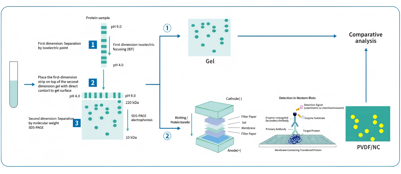
Figure 1: Workflow of antibody coverage verification via 2D-WB
HZSKBIO® has independently developed the Immunomagnetic Bead Separation (IMBS) technology[4]. Leveraging the semi-liquid state of immunomagnetic beads, the beads fully mix and bind with HCP samples under suspension conditions. Proteins remain undenatured throughout the workflow, and the binding mechanism mirrors ELISA testing conditions, delivering antibody coverage results that reflect authentic assay performance.
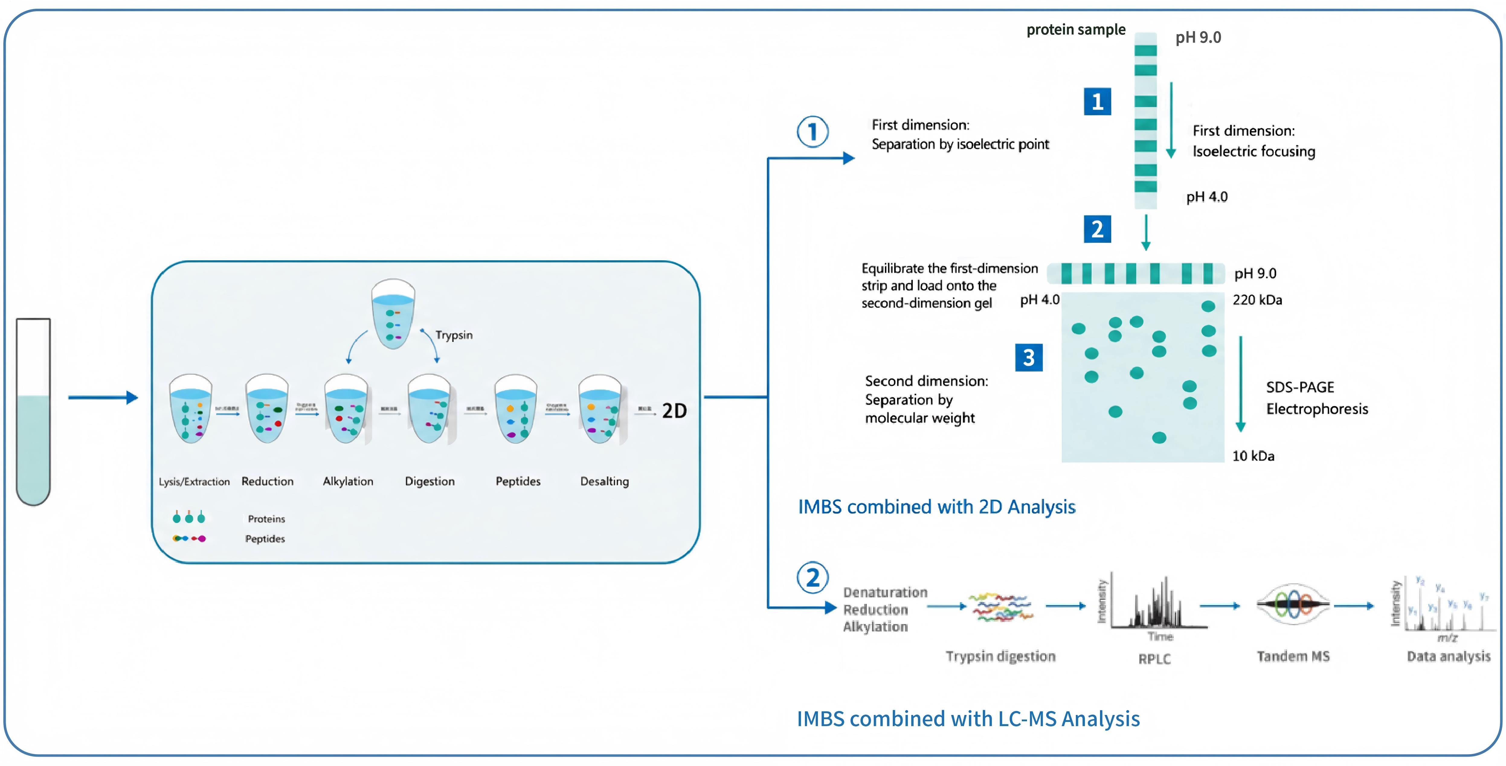
IMBS Technical Workflow (Figure 2)
Antibody coverage testing involves complex experimental workflows, requiring full validation of critical steps during method establishment to demonstrate assay robustness. HZSKBIO® has comprehensively validated key parameters during IMBS platform development, with validated study data reviewed by professional panels. Key validation items are summarized below:
Method Optimization: Fine-tune critical parameters including antibody-bead conjugation ratio, wash buffer volume, wash cycles and elution rounds to secure assay robustness;
In-process Quality Control: Two-dimensional electrophoresis consists of lengthy multi-step procedures with limited intermediate control. Fluorescent labeled peptides are integrated into isoelectric focusing (IEF) to rapidly evaluate IEF performance. 40 ng silver stain quality control markers are loaded on both sides of SDS-PAGE gels to standardize silver staining signal development;
Assay Repeatability: Repeatability verified for both 2D and LC-MS readouts. The 2D analysis achieves >80% repeatability, while LC-MS analysis reaches >90% repeatability;
Loading Mass Optimization: Confirm optimal sample loading volumes for 2D and LC-MS workflows to eliminate result bias induced by inconsistent loading amounts;
False Positive Elimination: Assess non-specific binding between samples and negative control antibodies to rule out false positive signals generated by IMBS procedure parameters.
* The full validation data set has been published in the Chinese Journal of New Drugs[5].
IMBS® Technology Platform Case Studies
CHO polyclonal antibody coverage was characterized using two IMBS-coupled analytical approaches:
IMBS-LC-MS (Figure 3A): Over 2,400 distinct proteins identified, with an overall CHO-HCP antibody coverage rate of 86.8%;
IMBS-2D (Figure 3B): Over 1,100 protein spots detected, with a coverage rate of 71.0%.
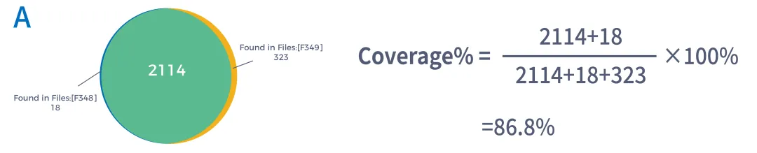
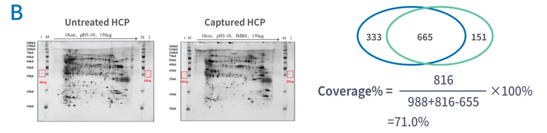
Figure 3 Antibody coverage analysis of CHO polyclonal antibodies using IMBS-LC-MS (Panel A) and IMBS-2D (Panel B), respectively
Over the one-year period spanning 2023 to 2024, HZSKBIO® has completed more than 50 antibody coverage testing projects with its fully validated coverage analysis platform operated under a compliant quality management system, supporting regulatory filings for various biological products. Detailed project data are summarized in the table below:
| Product Category | Client Background | Coverage Result | Application Scenario |
| Monoclonal Antibody / Bispecific Antibody / Fusion Protein | Leading domestic innovative biotech enterprise | 91.8% (LC-MS) | Post-market manufacturing process change |
| Monoclonal Antibody / Bispecific Antibody / Fusion Protein | Top-tier domestic oncology biotech | 85% (LC-MS) | BLA Submission |
| Monoclonal Antibody / Bispecific Antibody / Fusion Protein | Domestic ADC leader | 91.4% (LC-MS); 77.4% (2D) | BLA Submission |
| Recombinant Protein / Vaccine | Established domestic pharmaceutical enterprise | 91.4% (LC-MS) | BLA Submission |
| Recombinant Protein / Vaccine | Top domestic CDMO enterprise | 88% (LC-MS) | BLA Submission |
| Recombinant Protein / Vaccine | First-tier domestic biotech R&D enterprise | 88% (LC-MS) | Clinical Trial Stage |
| CGT Product | Leading domestic ophthalmic gene therapy enterprise | 87.7% (LC-MS); 70.6%–84.2% (2D) | Phase III Clinical Trial |
| CGT Product | Top domestic cell therapy enterprise | 65.3%–77.8% (2D) | BLA Submission |
References
[1] Qiu X, Xiang JZ, Zhao J, et al. Interpretation of the Technical Guidelines for Pharmaceutical Change Studies of Licensed Biological Products (Trial)[J]. China Food and Drug Administration Magazine, 2023(2):28-35.
[2] Wang X, Hunter AK, Mozier NM. Host cell proteins in biologics development: Identification, quantitation and risk assessment[J]. Biotechnology and Bioengineering, 2009.
[3] Chinese Pharmacopoeia Commission. Guidelines for Validation of Quantitative Analytical Methods for Biological Samples. Chinese Pharmacopoeia 2020 Volume IV, General Chapter 9012. Beijing: China Medical Science Press, 2020.
[4] United States Pharmacopeia. <1132> Residual Host Cell Protein Measurement in Biopharmaceuticals, USP 45-NF 40. Rockville, MD: United States Pharmacopeial Convention, 2022.
[5] Dou MH, Chen QY, Zhang HS, et al. Application of Immunomagnetic Bead Separation (IMBS) Technology in Host Cell Protein Antibody Coverage Analysis. Chinese Journal of New Drugs, 2024.