Products
Host Cell Residue Detection
Process Impurity Assay
Advnetitious Agent/Microorganism Testing
Pyrogen / Endotoxin Detection
Genetic Stability
Cell Line Characterization
Viral Titer Assay
Detection Equipment
Biochemical Reagents

Fully USP <1132> & EP 2.6.34 Compliant | Orthogonal IMBS®-2D& IMBS®-LC-MS Solutions.
Anti-host cell protein (HCP) antibody coverage assessment is a mandatory critical reagent characterization study for all biologic regulatory filings. This assessment characterizes the fraction of process-relevant HCPs bound by your ELISA anti-HCP polyclonal antibodies, mitigating false-negative release risks stemming from undetected HCP impurities. Our proprietary IMBS® orthogonal platform delivers two complementary testing workflows to fit early process development, method validation, and global IND/BLA submissions for mAbs, ADCs, recombinant proteins, and cell & gene therapy (CGT) products.
Global regulatory authorities — including FDA, EMA, USP, EP, ICH(Q2(R1),Q6B)— explicitly mandate full antibody coverage characterization for HCP immunoassays:
USP <1132> Residual Host Cell Protein Measurement in Biopharmaceuticals: Requires orthogonal testing to demonstrate antibodies recognize the majority of process-derived HCPs
EP 2.6.34 Host-Cell Protein Assays: Mandates coverage data to justify ELISA suitability for batch release
ICH & FDA Guidance: Orthogonal mass spectrometry readouts are strongly recommended over standalone 2D Western Blot (2D-WB) to avoid underreporting coverage from denaturation artifacts
Conventional 2D-WB relies on fully denatured HCPs during separation, which fails to mimic native ELISA binding conditions and often underestimates real antibody recognition performance. Our IMBS® technology solves this regulatory pain point via native suspension-phase immunocapture.
IMBS® covalently immobilizes anti-HCP polyclonal antibodies on magnetic microbeads for native-phase HCP capture, followed by two orthogonal analytical endpoints: IMBS®-2D Electrophoresis and IMBS®-LC-MS.
Standard IMBS® Workflow
1.Covalent antibody conjugation & bead blocking to reduce non-specific binding
2.Suspension incubation with process-mimicked HCP lysate (native, non-denaturing conditions matching ELISA)
3.Stringent wash steps to remove unbound HCP impurities
4.Low-pH elution of antibody-captured HCPs
5.Downstream orthogonal analysis: 2D gel imaging or high-resolution LC-MS identification
Core IMBS® Technical Superiorities vs Traditional 2D-WB / Resin AAE
Native ELISA-matching binding environment – no HCP denaturation, data reflects real ELISA performance
Superior enrichment & specificity – minimal non-specific adsorption compared to affinity resin columns
Dual orthogonal readouts: IMBS®-2D for visual spot coverage mapping; IMBS®-LC-MS for digital, quantifiable whole-proteome coverage
Proprietary SHENTEK® AbunProteoX kit depletes high-abundance therapeutic drug substance to eliminate MS signal interference
Regulatory & Quality System
ISO 13485 & CNAS accredited laboratory with full in-house method validation packages for global filing
Pre-built regulatory-ready study reports aligned with FDA/EMA IND/BLA documentation expectations
Orthogonal dual-method design fully satisfies USP/EP orthogonal testing requirements
Technical Flexibility
Two customizable workflows: IMBS®-2D for routine visual coverage screening; IMBS®-LC-MS for deep digital proteome characterization
Supports all mainstream expression systems: CHO, E. coli, HEK293, Vero, Sf9, and custom cell lines
Compatible with commercial off-the-shelf anti-HCP antibodies and client custom process-specific antibodies
Global Project Delivery Capacity
50+ annual HCP coverage projects covering mAb, ADC, recombinant protein, viral vector, and cell therapy pipelines
Proven regulatory submission track record for China, US, and EU dual filings
Dedicated regulatory scientific writing support for response to agency questions
Standard LC-MS HCP analysis suffers severe interference from overabundant therapeutic protein, masking low-level HCP peptides and distorting coverage results. Our proprietary SHENTEK® AbunProteoX Low Abundance Host Residual Protein Enrichment Kit selectively depletes target drug substance, drastically boosting detectable HCP peptide groups.
Key LC-MS Deliverables
Full inventory of all HCP species captured by your anti-HCP antibody (protein-level annotation)
Classification of high-risk HCPs (immunogenic, protease, degradation-inducing impurities)
Quantitative coverage percentage based on unique peptide groups
Actionable data to guide downstream purification process optimization and process validation risk assessment
SHENTEK® Enrichment Effect
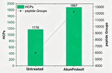
Figure. HCP peptide group counts identified from untreated vs. AbunProteoX (SHENTEK®) treated samples
| Product Modality | Client Profile | Testing Workflow | Regulatory Submission Purpose |
| Monoclonal antibody | Major biotech | IMBS®-LC-MS | Post-approval process change validation |
| Monoclonal antibody | Leading domestic oncology biotech | IMBS®-LC-MS | BLA |
| ADC Antibody Drug Conjugate | Premier ADC developer | IMBS®-2D | BLA |
| Recombinant protein | National biopharma R&D institute | IMBS®-LC-MS | BLA |
| CGT | National leader in ophthalmic gene therapy | IMBS®-LC-MS | Phase III clinical trial |
| CGT | Leading cell therapy enterprise | IMBS®-LC-MS | BLA |
*Representative partial project portfolio
| Testing Workflow | Sample Criteria (Process-Mimicked HCP Lysate, Bradford Quantified) | Typical Antibody Input | Standard Project Cycle (working days) |
| IMBS®-2D | Total protein ≥60 mg; Concentration ≥2 mg/mL | 1 mg polyclonal anti-HCP antibody | 40 WD |
| IMBS®-LC-MS | Total protein ≥60 mg; Concentration ≥4 mg/mL | 1 mg polyclonal anti-HCP antibody | 35 WD |
Supplementary Sample Guidance
Samples must be representative of your full manufacturing harvest/process stream
Frozen lysate shipped on dry ice; avoid repeated freeze-thaw cycles to prevent HCP degradation
Provide blank null cell lysate reference material for full proteome background mapping
Full study report with raw chromatogram/gel imaging data
Digital LC-MS dataset: Excel list of all antibody-recognized HCPs, peptide counts, risk classification
Coverage percentage summary (2D spot ratio / LC-MS unique protein ratio)
Regulatory-ready appendix for IND/BLA submission (method validation summary, IMBS platform qualification data)
Visual 2D gel comparison maps (total HCP vs antibody-captured HCP spots)
Q1: What typical coverage percentages do you observe across past projects?
A: Most commercial generic anti-HCP antibodies show LC-MS coverage higher than 80%, with numerous batches hitting above 90%. The majority of our custom process-specific antibodies reach ≥85% coverage via IMBS®-LC-MS, though results differ depending on cell host and immunization design.
Q2: Is a higher coverage percentage always preferable for regulatory filings?
A: Assuming rigorous, artifact-free testing, higher coverage is universally favorable as it demonstrates broader antibody recognition of your process HCP pool. Method baseline thresholds vary by technique:
· IMBS®-2D visual spot coverage: Industry & regulatory acceptable baseline ≥70%
· IMBS®-LC-MS digital proteome coverage: Standard filing expectation ≥80% The higher LC-MS threshold stems from superior mass spec sensitivity and quantitative accuracy compared to gel-based 2D imaging.
Q3: What is the regulatory acceptable minimum coverage threshold for BLA/IND?
A: Regulators do not enforce an absolute hard cutoff, but global industry consensus aligned with USP <1132> guidance:
· 2D gel-based coverage: ≥70% minimum justification threshold
· Orthogonal LC-MS coverage: ≥80% preferred for full regulatory confidence Coverage below these benchmarks requires extensive scientific justification to prove no high-risk unrecognized HCPs exist in your process.
Beyond coverage percentage, regulators increasingly expect manufacturers to assess the immunogenic risk of individual HCP species — particularly those with known biological activity (e.g., proteases, cytokines) — that may evade detection.
Q4: How does IMBS® differ from traditional 2D-WB coverage testing?
A: Conventional 2D-WB fully denatures HCPs during electrophoresis, disrupting native antibody-antigen epitopes and artificially lowering measured coverage. IMBS® performs HCP capture in native suspension matching ELISA liquid-phase conditions, delivering biologically relevant coverage data that directly correlates with your routine release assay. It also reduces non-specific background compared to static affinity resin chromatography.
Q5: Can I submit only my commercial off-the-shelf HCP antibody for coverage testing?
A: Yes, we support coverage characterization for all commercial CHO/E. coli/HEK HCP antibody kits. We recommend pairing IMBS-LC-MS orthogonal testing to identify process-specific HCPs missed by generic antibodies, a common FDA inspection observation point.
Q6: What if my program is cell & gene therapy (viral vector/cell therapy)? Are there special testing adjustments?
A: Our IMBS platform is fully optimized for CGT matrix complexity (high host cell debris, low HCP abundance). The SHENTEK® AbunProteoX enrichment kit removes viral vector/cell product interference to recover low-level residual HCPs, and we have dedicated CGT case studies for Phase III and BLA filings.
Establishing a robust in-house HCP coverage characterization capability is technically feasible, but it requires significant capital investment, specialized expertise, and extensive method development time — resources that many drug development teams prefer to allocate to core pipeline activities.
The Hidden Costs of In-House Development
| Cost Factor | What It Entails |
| Instrumentation | High-resolution LC-MS/MS systems, 2D electrophoresis rigs, imaging systems, and supporting infrastructure — representing a $500K–$1M+ capital outlay |
| Expertise & Training | Recruiting or upskilling scientists with deep proteomics and biopharma regulatory experience — a scarce and costly talent pool |
| Method Development | 6-12 months of internal effort to develop, qualify, and document a fit-for-purpose HCP coverage method |
| Regulatory Risk | Unfamiliarity with evolving FDA/EMA expectations can lead to review delays, additional questions, or even rejection of your coverage strategy |
返回