Products
Host Cell Residue Detection
Process Impurity Assay
Advnetitious Agent/Microorganism Testing
Pyrogen / Endotoxin Detection
Genetic Stability
Cell Line Characterization
Viral Titer Assay
Detection Equipment
Biochemical Reagents

When testing complex biological samples, monocyte cell lines with high homogeneity deliver superior detection stability compared to mixed peripheral blood mononuclear cells (PBMCs), exhibiting greater tolerance to fluctuations in complex sample matrices and culture environments.
How does monocyte activation test (MAT) cell incubation specifically recognize pyrogen-related molecules without interference from sample matrices? Toll-Like Receptors (TLRs) play an indispensable role.
01 Accurate TLR Profiling: The Core to Overcoming MAT Application Limitations
TLRs are transmembrane receptor proteins on immune cell surfaces that recognize pathogens[1], widely distributed on cell membranes and endomembrane structures. This "receptor army" consists of 10 functional subtypes that coordinate to comprehensively identify exogenous threats.
Each subtype features highly targeted distribution: TLR1, 2, 4, 5, 6 localize on the cell plasma membrane, while TLR3, 7, 8, 9 reside within endosomes[2]; certain subtypes can shuttle flexibly between the cell membrane and endosomes. This precise compartmentalization enables each subtype to perform dedicated functions and specifically recognize distinct exogenous pyrogens (pathogen-associated molecular patterns, PAMPs) (see Table 1[3]).
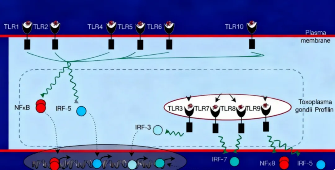
Table 1 List of TLR Subtypes and Corresponding Captured PAMPs
| TLRs | PAMPs |
TLR1 | Bacteria (Mycobacteria, Neisseria, spirochetes, triacylated lipopeptides) |
TLR2 | Bacteria, peptidoglycan, muramic acid (lipoteichoic acid), yeast zymosan, lipoproteins, glycolipids, heat shock proteins, defensins |
TLR3 | Viral double-stranded RNA (dsRNA) and polyinosinic-polycytidylic acid (Poly-IC) |
TLR4 | Lipopolysaccharide (LPS) from Gram-negative bacteria, endogenous ligands including heat shock proteins, fibronectin, heparan sulfate, hyaluronic acid, synthetic Taxol |
TLR5 | Bacterial flagellin |
TLR6 | Acylated lipopeptides |
TLR7 | Viral single-stranded RNA (ssRNA, e.g., influenza virus, HIV) and synthetic small-molecule antiviral analogs |
TLR8 | Viral ssRNA and synthetic pharmaceutical compounds |
TLR9 | Unmethylated CpG motifs from bacterial DNA |
TLR10 | Ligand unknown |
In short, the immune response of MAT monocyte cell lines to pyrogens entirely relies on TLRs—the cellular "scout troop". Specific binding between exogenous pyrogens and matching TLRs initiates a cascade of immune responses.
02 24-Hour Immune Response: A Microscopic Signal Relay Race
The 24-hour co-incubation of monocytes with test samples during MAT testing constitutes the critical phase of immune signal transduction.
Upon binding PAMPs, TLRs undergo homodimerization; their intracellular TIR domains act as "immune signal checkpoints" and rapidly recruit the adaptor protein Myeloid Differentiation Primary Response Protein 88 (MyD88).
Following recognition by MyD88, this primary relay initiates downstream signal transmission via ubiquitination cascades[3].
The core signal effector IKK complex receives the signal and phosphorylates IκB, the inhibitory factor of NF-κB, thereby releasing NF-κB.
The terminal messenger NF-κB translocates into the nucleus[3] and activates transcription of inflammatory factor genes, driving expression of endogenous pyrogen cytokines including TNF-α, IL-1β and IL-6[4].
This precisely coordinated immune signaling cascade completes the cellular defense against inflammation and infection, ultimately triggering physiological responses such as fever. These cytokines serve as the core pyrogen biomarkers measured in MAT assays.
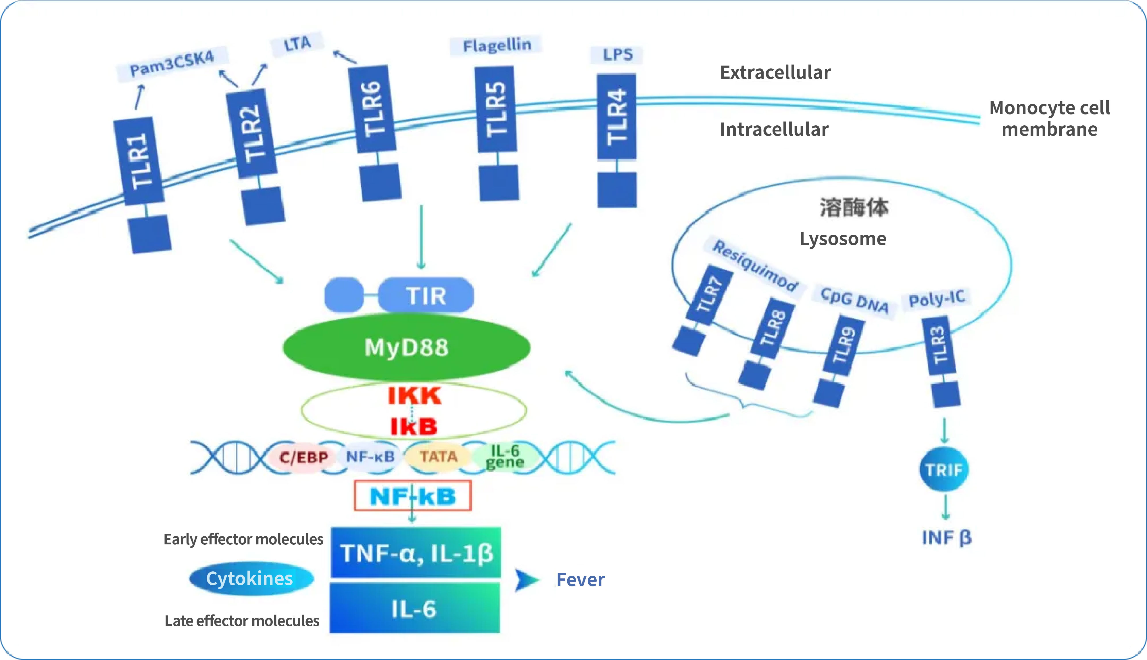
Figure 2 Schematic diagram of immune signal transduction cascades
03 Accurate TLR Profiling: Initiating the First Step of Monocyte Immune Response
Monocyte cell lines supplied in the PyroSHENTEK™ Pyrogen Detection Kit undergo systematic validation of TLR transmembrane proteins via Western blot analysis (Figure 3).
This process constructs a full-spectrum cellular sensing network. Equipped with a complete TLR expression profile, MAT cells fully cover recognition of all pyrogen types, laying a solid foundation for robust detection performance from the source.
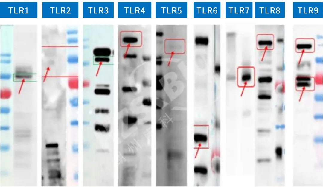
Figure 3 TLR expression profiling results of MAT cells from PyroSHENTEK™
Cellular responsiveness to pyrogens heavily depends on reaction speed and efficiency. Relevant research provides key theoretical evidence confirming that monocyte differentiation induced by ligands such as LPS exhibits clear concentration- and time-dependent characteristics[2].
Based on extensive literature research and in-depth analysis of immune response mechanisms, HZSKBIOⓇ optimized and finalized critical differentiation induction parameters for MAT cells, markedly improving cellular responsiveness in complex sample matrices (sample applicability data shown in Table 2). The cells also display robust reactivity to non-endotoxin pyrogens across varying concentrations (results shown in Figure 4).
Table 2 Sample Applicability of HZSKBIOⓇ MAT Assay
| Drug/Raw Material Category | Representative Products | Detection Kit | Preprocessing Kit | ||||
| Monoclonal Antibodies | Rituximab | Sintilimab | Adalimumab | Nimotuzumab | Ranibizumab | MAT Kit | / |
Etanercept | Ruxolitinib | Bevacizumab | Evolocumab | Alirocumab | |||
Vaccines | Human Rabies Vaccine | Lyophilized Inactivated Hepatitis A Vaccine | MMR Combined Live Attenuated Vaccine | Quadrivalent Influenza Split Vaccine | Group A/C Meningococcal Vaccine | ||
Influenza Split Vaccine | Live Attenuated Varicella Vaccine | ||||||
Recombinant Proteins | Recombinant Insulin Injection | Teriparatide Injection | Recombinant Human Erythropoietin Injection | Recombinant Human Interferon α-2b Injection | Recombinant IL-11 for Injection | ||
Glargine Insulin | |||||||
Blood Products | Human Prothrombin Complex Concentrate | Intravenous Immunoglobulin | Human Coagulation Factor VIII | Human Albumin | |||
Biochemical Pharmaceuticals | Urokinase | Succinylated Gelatin | Collagenase | Ademetionine 1,4-Butanedisulfonate | Glutathione | ||
| Ziprasidone Mesylate | Indocyanine Green | Mannatide | Nicotinamide | Reduced Glutathione | |||
Adrenaline Hydrochloride | |||||||
Injectable Herbal Preparations | Shenmai Injection | Xiangdan Injection | Normal Saline | ||||
Raw & Auxiliary Materials | BSA | Fetal Bovine Serum | Cefotaxime Sodium | Troxerutin | |||
Antibiotics | Cefotaxime Sodium | Tedizolid Phosphate | Famotidine | Palonosetron Hydrochloride | Azlocillin Sodium | ||
Cefuroxime Sodium | Amikacin Sulfate | ||||||
Small-Molecule Drugs | Polymyxin B & E | MAT Kit | Preprocessing Reagent A | ||||
Epirubicin Hydrochloride | MAT Kit | Preprocessing Reagent B | |||||
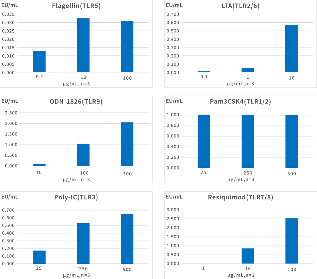
Figure 4 Response profiles of PyroSHENTEK™ MAT cells to various non-endotoxin pyrogens (NEPs) across different concentrations
04 PyroSHENTEK™ Pyrogen Detection Kit (MAT Assay)
Compliant Traceability Foundation: Cell lines are commercially licensed with fully traceable supply chains, fully meeting regulatory filing requirements and mitigating compliance risks at the source.
Multi-Dimensional Optimization for Broad Application Scope: Dual optimization strategies covering cellular performance enhancement and sample preprocessing upgrades continuously break detection limitations and expand applicable sample ranges.
Stable and Reliable Quality: Subjected to comprehensive performance verification and dual certification by regulatory authorities and end customers, delivering consistent, reproducible test results.
Proven Industrial Application: Successful regulatory filing cases in the EU market translate laboratory scientific optimizations into practical, implementable testing solutions.
References
[1] Kaczmarek, A., et al. (2020) ‘Expression of surface and intracellular Toll-like receptors by mature mast cells’, PubMed. Available at: https://pubmed.ncbi.nlm.nih.gov/28450795/ (Accessed: 24 October 2025)
[2] Mita, Y., Dobashi, K., Nakazawa, T. and Mori, M. (2001) ‘Induction of Toll-like receptor 4 in granulocytic and monocytic cells differentiated from HL-60 cells’, British Journal of Haematology, 112(4), pp. 1041–1047. doi: 10.1046/j.1365-2141.2001.02658.x. Erratum in: British Journal of Haematology, 115(3), p. 715. PMID: 11298604
[3] Blasius, A.L. and Beutler, B. (2010) ‘Intracellular toll-like receptors’, Immunity, 32(3), pp. 305–315. doi: 10.1016/j.immuni.2010.03.012. PMID: 20346772
[4] Tanaka, T., Narazaki, M., Masuda, K. and Kishimoto, T. (2016) ‘Regulation of IL-6 in Immunity and Diseases’, Advances in Experimental Medicine and Biology, 941, pp. 79–88. doi: 10.1007/978-94-024-0921-5_4. PMID: 27734409